Estimated reading time: 4 minute
Table of Contents
OpenStax College, CC BY 3.0 https://creativecommons.org/licenses/by/3.0, via Wikimedia Commons
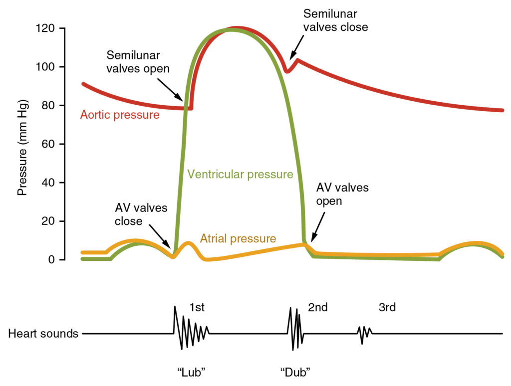
What are the heart sounds heard during auscultation?
There is an important difference between heart sounds and heart murmurs. Heart sounds, are physiological sounds that are generally considered normal, but heart murmurs, are abnormal sounds heard due to blood turbulence and may be a sign of a disease.
In small animals (cats, dogs) it is usually only 2 heart sounds (S1 and S2) are heard. However, in horses, in addition to these sounds, 3rd and 4th heart sounds (S3 and S4) It can also be heard. In cats and dogs, the 3rd and 4th sounds are usually heard a pathological condition points out.
What are the definitions of heart sounds and their formation mechanisms?
1. First Heart Sound (S1)
• Formation Mechanism: Closure of the mitral and tricuspid valves (AV valves) It consists of.
• Sound Place: Closure of the left and right atrioventricular (AV) valves creates this sound.
• Physiological Importance: The S1 sound indicates the beginning of the heart's contraction (systole) phase. This sound is known as the "lub" sound and is usually strong in tone.
Clinical Significance: Changes in the S1 sound may indicate a problem with the AV valves (mitral and tricuspid). If this sound is if weak, pericardial effusion, obesity or pneumothorax come to mind. If the sound more strong may cause hyperdynamic circulation (fever, anemia, etc.).
2. Second Heart Sound (S2)
• Formation Mechanism: Closure of the aortic and pulmonary valves (semilunar valves) It consists of.
• Sound Place: It occurs when the valves close as the blood from the left and right ventricles passes into the aorta and pulmonary artery.
• Physiological Importance: The S2 sound indicates the point at which the heart's contraction (systole) phase ends and the relaxation (diastole) phase begins. This sound is known as the "dub" sound.
Clinical Significance: Changes in the S2 sound may indicate a problem with the aortic and pulmonary valves. For example, in the case of pulmonary hypertension S2 sound is split This split is associated with the aortic and pulmonary valves not closing simultaneously. Pulmonary stenosis or pulmonary hypertension In some cases this split sound can be heard clearly.
3. Third Heart Sound (S3)
• Formation Mechanism: Ventricular wall vibration This vibration occurs during rapid filling of the ventricle.
• Sound Place: Usually heard in the left ventricle.
• Physiological Importance: The S3 sound is produced by vibration of the ventricular wall during rapid ventricular filling. It is usually not heard in healthy small animals.
Clinical Significance: S3 sound, usually with cardiomyopathies This sound is caused by the hardening of the ventricular wall and the inability of the ventricle to expand sufficiently. Specifically, dilated cardiomyopathy (DCM) This sound becomes more pronounced in cases of S3 sound, heart failure It may be an early symptom. Therefore, when the S3 sound is noticed, echocardiography The thickness and functionality of the ventricular wall should be checked.
4. Fourth Heart Sound (S4)
• Formation Mechanism: The forced expulsion of blood into the ventricle during atrial contraction This sound is an indication of a problem with the relaxation process of the ventricle.
• Sound Place: Usually heard in the left ventricle.
• Physiological Importance: The S4 sound is produced by the pushing of blood into the ventricle during atrial contraction. Normally, this sound is not heard, but when there is a problem with ventricular relaxation (for example, hypertrophic cardiomyopathy, HCM) this sound becomes clear.
Clinical Significance: Hypertrophic cardiomyopathy (HCM), is a heart disease that is especially common in cats. In this disease, S4 sound can be heard due to thickening of the ventricular muscle. S4 sound also in old cats may be heard because ventricular flexibility may decrease with age. The S4 sound is an early indicator of difficulty in ventricular filling. In this case, echocardiography should be performed and ventricular thickness should be assessed.
How is clinical evaluation of heart sounds made?
S1 (First Heart Sound)
• Mechanism: Closure of mitral and tricuspid valves
• Clinical Significance: It is the beginning of the heart's contraction (systole) phase. Changes may indicate mitral and tricuspid valve diseases.
S2 (Second Heart Sound)
• Mechanism: Closure of aortic and pulmonary valves
• Clinical Significance: It is the beginning of the relaxation (diastole) phase of the heart. Split S2 may indicate pulmonary hypertension.
S3 (Third Heart Sound)
• Mechanism: Ventricular pulsation (rapid filling)
• Clinical SignificanceHeard in conditions where ventricular dilatation is present, such as dilated cardiomyopathy (DCM) and heart failure.
S4 (Fourth Heart Sound)
• Mechanism: Atrial contraction
• Clinical Significance: Associated with hypertrophic cardiomyopathy (HCM) and impaired ventricular relaxation in older cats.
Other Articles You May Be Interested In
“Doubt is the beginning of knowledge.”
René Descartes
