Estimated reading time: 8 minute
Table of Contents
The classification of cardiac arrhythmias seen in cats and dogs is as follows.
Supraventricular Arrhythmias
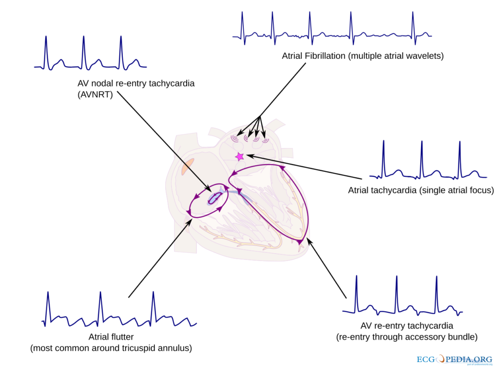
They are rapid and irregular rhythm disorders originating from the atria or atrioventricular node, where the heart rate per minute is within or above the reference ranges that vary according to species, gender and age.
CardioNetworks: Drj, CC BY-SA 3.0 https://creativecommons.org/licenses/by-sa/3.0, via Wikimedia Commons
• Sinus tachycardia: It is a regular arrhythmia originating from the sinus node, in which the heart rate exceeds the reference range for age, species and gender.
• Atrioventricular nodal reciprocating tachycardia: Rapid and regular heartbeats occur due to reentrant electrical activity circulating within the AV node.
• Focal junctional tachycardia: It is a rapid and regular rhythm disturbance originating from a specific point around the AV node.
• Non-paroxysmal junctional tachycardia: It is a type of tachycardia that is sustained and slow-onset and originates from foci around the AV node.
• Atrioventricular reciprocating tachycardias: They are fast rhythm disorders that occur due to electrical activity circulating between the AV node and this pathway in cases where there is an extra conduction pathway.
Orthodromic atrioventricular reciprocating tachycardia: The electrical impulse travels from the AV node to the ventricles via the normal pathway, but returns to the atrium via the accessory pathway.
Antidromic atrioventricular reciprocating tachycardia: The electrical impulse travels to the ventricles via the accessory pathway and returns to the atrium via the AV node.
• Persistent junctional reciprocating tachycardia: It is a type of reentrant tachycardia that is constantly present and circulates around the AV node.
• Focal atrial tachycardia: It is a rapid and regular rhythm disturbance originating from a specific area of the atrium.
• Multifocal atrial tachycardia or chaotic atrial rhythm: It is an irregular and rapid rhythm originating from multiple foci in the atrium.
• Macroreentrant atrial tachycardia: It is a type of tachycardia that produces a wide reentrant loop in the atria.
Cavo-tricuspid isthmus dependent atrial flutter: It is a rhythm disorder that creates a loop between the tricuspid valve in the right atrium and the inferior vena cava.
Cavo-tricuspid isthmus independent atrial flutter: It is a macroreentrant atrial tachycardia that develops outside the cavo-tricuspid isthmus region.
• Atrial fibrillation: It is a rhythm disorder in which the ventricles are stimulated irregularly due to irregular and rapid contractions of the atria.
Ventricular Arrhythmias
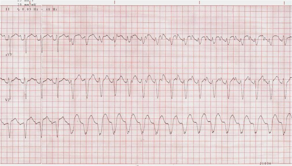
They are irregular or rapid rhythm disorders originating from the ventricles, where the heart rate per minute may be below, within or above the reference ranges that vary according to species, gender and age.
CardioNetworks: Giniger, CC BY-SA 3.0 https://creativecommons.org/licenses/by-sa/3.0, via Wikimedia Commons
• Idioventricular rhythm: Heart rate per minute originating from the ventricles below the reference ranges that vary according to species, gender and age It is a regular rhythm and is usually seen in cases where the AV node is dysfunctional.
• Accelerated idioventricular rhythm: It is a generally transient and benign rhythm disorder in which the rate of the idioventricular rhythm increases and is within or above the reference ranges that vary according to species, gender and age.
• Monomorphic ventricular tachycardia: It is a regular, rapid rhythm originating from the ventricles and seen with a fixed QRS morphology. Heart rate per minute Above reference ranges that vary by species, gender and age.
Unmaintained: It is a monomorphic ventricular tachycardia that lasts less than 30 seconds and terminates spontaneously.
Recurrent: It is a ventricular tachycardia that recurs in short episodes.
Sustained: Monomorphic ventricular tachycardia that lasts longer than 30 seconds or requires intervention.
Continually: It is a type of tachycardia that continues continuously and usually causes hemodynamic disturbances.
• Ventricular flutter: It is a regular but life-threatening rhythm in which QRS waves merge to form a sinusoid due to very rapid ventricular impulses. The heart rate per minute is usually It is well above the reference ranges that vary according to species, gender and age..
• Polymorphic ventricular tachycardia: It is an irregular rapid rhythm originating from the ventricles and in which the QRS complexes are variable in shape and amplitude. The heart rate is usually Above reference ranges that vary by species, gender and age.
• Torsades de pointes: It usually develops on the basis of QT prolongation, where the amplitude and direction of QRS complexes fluctuate. Above reference ranges that vary by species, gender and age is a ventricular tachycardia.
• Bidirectional ventricular tachycardia: It is a rhythm originating from the ventricles in which successive QRS complexes change direction along different axes. Heart rate per minute Above reference ranges that vary by species, gender and age.
• Ventricular fibrillation: A lethal rhythm in which the ventricles fail to produce effective cardiac output due to irregular, rapid, and unsynchronized contractions. Heart rate per minute It is well above the reference ranges that vary according to species, gender and age..
Bradycardia and Conduction Disorders
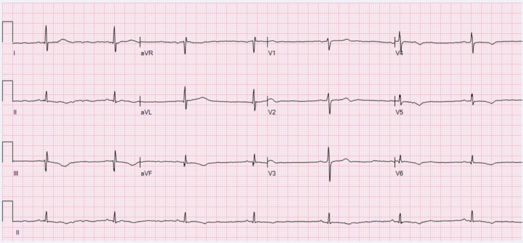
Rhythm and conduction problems characterized by slowing, interruption or irregularity in the production of electrical impulses by the heart or in the conduction of these impulses.
Ewingdo, CC BY-SA 4.0 https://creativecommons.org/licenses/by-sa/4.0, via Wikimedia Commons
• Sinus bradycardia: Heart rate originating from the sinus node and increasing per minute below the reference ranges that vary according to species, gender and age It is a regular rhythm.
• Sinus arrest: It is an arrhythmia in which the heart pauses and does not beat for a while due to the temporary stoppage of the sinus node.
• Sinus arrest: It is a serious conduction disorder in which the heart beats stop due to the failure of the sinus node to produce electrical impulses.
• Atrial arrest: It is a disorder that causes the heart to beat irregularly because the atria stop producing electrical activity.
• Sino-ventricular rhythm: It is a rare rhythm in which the signals from the sinus node are transmitted directly to the ventricles without passing to the atria.
• Asystole or ventricular arrest: It is a fatal condition in which the ventricles of the heart completely stop electrical and mechanical activity.
• Pulseless electrical activity (PEA) or electromechanical dissociation: A condition in which electrical activity is seen on the ECG, but no pulse is detected and there is no effective cardiac output.
• Intra-atrial conduction delay or block: It is a conduction disorder in which electrical conduction within the atrium is slowed down or completely interrupted.
• Sino-atrial block: It is a conduction disorder in which electrical signals from the sinus node cannot be transmitted to the atrium.
• Atrioventricular block: They are conduction disorders caused by a delay or complete interruption of electrical conduction from the atria to the ventricles.
First degree: It is a block in which conduction in the AV node is delayed but not completely interrupted.
Second degree: It is a block in which some of the atrial impulses cannot be transmitted to the ventricles.
Wenckebach (Mobitz Type 1): A condition in which the PR interval becomes progressively longer and eventually a ventricular beat fails to occur.
Mobitz Type 2: It is a more severe block in which some atrial impulses fail to conduct with fixed PR intervals.
Fixed 2:1: A condition in which one out of every two atrial impulses is conducted to the ventricles.
Advanced degree: It is a condition in which several consecutive atrial impulses fail to be conducted to the ventricles.
Third degree (Complete block): It is a conduction disorder in which atrial and ventricular activities are completely separated.
Paroxysmal: It is a temporary AV block that occurs in attacks.
• Intraventricular blocks: These are blocks in which the electrical conduction between the ventricles is partially or completely blocked.
Monofascicular: A single bundle branch block (right bundle branch block or left bundle branch block) is seen.
Bifascicular: It is a block in which two branches (right branch and left anterior or posterior branch) are affected.
Trifascicular: It is a serious conduction disorder in which three branches are affected.
Ectopic Beats and Rhythms
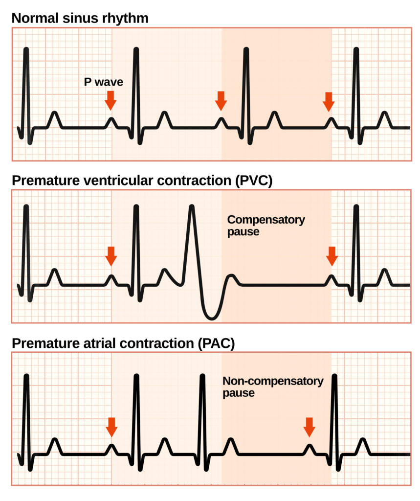
They are abnormal impulses and irregular rhythms originating from ectopic foci such as the atrium, ventricle or AV node, outside the normal electrical conduction pathway of the heart.
YitzhakNat, CC BY-SA 4.0 https://creativecommons.org/licenses/by-sa/4.0, via Wikimedia Commons
• Supraventricular ectopic beats and rhythms: They are premature or abnormal rhythms and beats originating from supraventricular regions such as the atrium or atrioventricular node, originating from a focus other than the normal sinus node.
Atrial ectopic beats: They are premature and irregular heartbeats originating from a focus other than the normal sinus node of the atrium.
Atrial ectopic rhythm: It is a regular but abnormal rhythm originating from a focus in the atrium other than the sinus node.
Junctional ectopic beats: They are premature and irregular heartbeats originating from the AV node region.
Junctional ectopic rhythm: It is a regular but abnormal rhythm originating from a focus around the AV node.
Atrial parasystole: They are continuous and irregular beats produced by a focus in the atrium independent of the sinus node.
Dissociation or unilateral atrial rhythm: It is an irregular rhythm that originates in one part of the atrium and progresses independently of other parts.
• Ventricular ectopic beats and rhythms: They are premature, irregular or independent rhythms and beats originating from the ventricles and originating from a focus outside the normal conduction pathway.
Ventricular ectopic beats: They are irregular heartbeats characterized by premature and often wide QRS complexes originating in the ventricles.
Idioventricular rhythm: It is a slow and regular rhythm originating from the ventricles when the sinus node or AV node is dysfunctional.
Ventricular parasystole: They are irregular and continuous beats that originate from a focus in the ventricle and occur independently of the normal rhythm of the ventricle.
Other Articles You May Be Interested In
“A sufficiently advanced technology is indistinguishable from magic.”
Arthur C. Clarke,en
