Estimated reading time: 10 minute
Table of Contents
- Definition and History
- Use in Humans
- Advances in Veterinary Cardiology
- Clinical Studies
- Eligibility Criteria for the Startup
- Diagnostic Criteria for Severe Mitral Regurgitation (MR)
- Evaluation Methods
- Mitral Functional Anatomy and Its Relationship to Success
- Final Assessment for Edge-to-Edge Repair
- Transcatheter Edge-to-Edge Mitral Valve Repair (TEER)
- Surgical Intervention
- Viewing and Viewing Angles
- Anticoagulation
- V-Clamp Measurement and Placement
- V-Clamp Dimensions:
- Opening the Heart Hole and Preparing the Entry Way:
- Mitral Valve Passage:
- V-Clamp Placement:
- Mitral Valve Leaflet Capture:
- Control and Evaluation
Definition and History
Edge-to-edge mitral valve repair The concept was first developed by a cardiac surgeon Ottavio Alfieri by, to correct prolapse (prolapse) of the anterior leaflet of the mitral valve It has been proposed as a surgical solution for this technique. the prolapsed leaflet is stitched to the leaflet opposite it A two-orifice (double-orifice) mitral valve is created.
Following this successful surgical repair procedure, transcatheter edge-to-edge mitral valve repair (TEER) technique was developed. This technique, a noninvasive approach has become a common treatment method in human patients.
Use in Humans
The TEER procedure in humans MitraClip device is done using.
• EVEREST feasibility studyThis is the first study to evaluate the efficacy and safety of the MitraClip device.
• EVEREST II and COAPT Two major studies titled, has proven the effectiveness of TEER in the treatment of severe mitral regurgitation (MR) and has led to its widespread acceptance.
• TEER in humans is no longer Safe and effective treatment option for severe mitral regurgitation is widely used as.
Advances in Veterinary Cardiology
The success achieved in humans led to the development of a similar device in veterinary medicine, designed specifically for dogs. Canine Mitral V-Clamp device.
Canine Mitral V-Clamp Device
• This device, Developed to treat degenerated mitral valve disease (DMVD) in dogs.
• Firstly, In 1 dogs with stage B8 degenerated mitral valve disease In a preliminary study, the feasibility and effectiveness of the device were demonstrated.
Clinical Studies
• In dogs with severe mitral regurgitation (MR) Prospective feasibility trial of the V-Clamp device has been carried out.
• The purpose of this study was to evaluate how safe and effective the device is in dogs.
• Transcatheter edge-to-edge mitral valve repair (TEER), Firstly a procedure developed in humansand MitraClip device It is widely used to treat severe mitral regurgitation (MR).
• In veterinary medicine, Canine Mitral V-Clamp device, similarly in dogs degenerate mitral valve disease (DMVD) is designed to treat.
• Preliminary studies, the use of this device in dogs demonstrates that it is applicable and safe.
• Wider applicability and effectiveness of the device, Prospective feasibility study in dogs with severe mitral regurgitation is evaluated with.
These developments in veterinary cardiology emergence of new, noninvasive options for the treatment of degenerated mitral valve disease It provides.
Eligibility Criteria for the Startup
Degenerative mitral regurgitation (DMR) Dogs that will be subjected to intervention due to American Society of Echocardiography (ASE) criteria for severe mitral regurgitation (MR) in humans must meet the adapted criteria.
• Dogs in ACVIM stage B2 or Cis considered a candidate for mitral intervention.
• Late stage D or dogs with atrial fibrillation (AF), for mitral intervention are not considered suitable candidates.
Diagnostic Criteria for Severe Mitral Regurgitation (MR)
To determine the severity of MR More than one criterion needs to be evaluated. Criteria supporting severe MR It is as follows:
• Holosystolic, eccentric colored current jets affecting the wall or the central colored stream jet area is greater than 50%
• E-wave dominant mitral inflow velocity ≥1.0 m/s
• Intense holosystolic triangular regurgitation profile on continuous wave Doppler
• Regurgitation fraction (RF) ≥50% (Proximal Isovelocity Surface Area (PISA) can be measured by volumetric or Doppler)
• Regurgitation volume (RVol) ≥1.0 mL/kg
Evaluation Methods
• Color Flow Doppler Evaluation:
• In different systolic images ve appropriate gain and Nyquist settings should be evaluated with.
• Assessing MR severity with a single image may incorrectly make the MR appear more severe than it actually is.
• Regurgitation Fraction (RF) Measurement:
• RF, PISA, Simpson volumetric and/or Doppler methods measurable.
• Measuring RF with multiple methods, increases the reliability of the evaluation.
• Vena Contracta (VC) Width, Regurgitant Volume (RVol) and Effective Regurgitant Orifice Area (ERO):
• These measurements can be taken into account, but varies depending on the size of the patient.
• The exact limit values for dogs have not yet been determined..
• Left Ventricle (LV) and Left Atrium (LA) Enlargement:
• Left ventricular diastolic internal diameter (LVIDdN) >1.9 within the higher education institution
• Left atrium/aortic root ratio (LA:Ao) >2.0 within the higher education institution
• These changes occur as a result of MRI. are secondary changes ve chronicity of the disease ve to the treatment applied It depends.
• Systolic Function Indices:
• Left ventricular ejection fraction (LVEF) <50% within the higher education institution
• Left ventricular end diastolic diameter (LVIDsN) >1.0 within the higher education institution
• These parameters may be an indication that the potential for reduction of MR is weak.
Mitral Functional Anatomy and Its Relationship to Success
• Mitral valve Functional anatomy, in the TEER procedure is one of the most important determinants of success and results.
• Dimensions of available dog devices Designed in 14 mm, 16 mm and 18 mm.
• Apical inflow-outflow echocardiographic image, The anterior-posterior (AP) mitral annulus diameter in mid-systole is between 14-20 mm. be recommended.
Final Assessment for Edge-to-Edge Repair
• Functional anatomical structure should be evaluated with transesophageal echocardiography (TEE) before the procedure..
• TEE more clearly demonstrates the size, anatomy and suitability for the procedure of the mitral valve.
1. What Dogs Are Eligible for TEER?
• Dogs in ACVIM B2 and C stages are the most suitable candidates.
• Dogs in Stage D ve dogs with atrial fibrillation (AF)are not eligible candidates for TEER.
2. Criteria for Assessing the Severity of MR
• Criteria such as holosystolic colored flow jets, regurgitation fraction (RF), vena contracta width and regurgitation volume (RVol) are used.
• Instead of a single image, evaluation with more than one method recommended.
3. Evaluation of Functional Anatomical Structure
• In dogs with appropriate anatomical structure (green category) the procedure has a higher success rate.
• Dogs of moderately fit constitution (yellow category) may benefit from the procedure, but the risks are greater.
• Dogs with unsuitable anatomical structure (red category) They are not eligible candidates for TEER.
4. Transesophageal Echocardiography (TEE) Evaluation
• TEE, is the most important tool in the final evaluation and decision-making phase.
• TEE, provides accurate assessment of the morphology, size and structure of the mitral valve.
In this process, correct patient selection, functional anatomical structure ve pre-procedural evaluation are the basic elements that directly affect success. Making the right assessment, increases the likelihood of the procedure being successful and the patient's quality of life.
Transcatheter Edge-to-Edge Mitral Valve Repair (TEER)
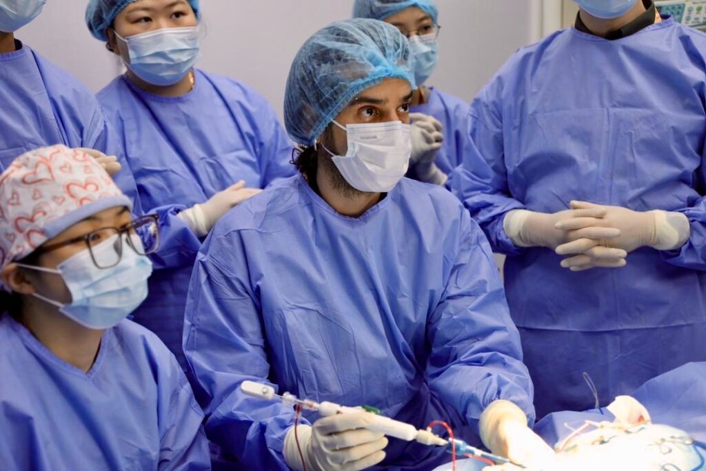
TEER procedure in dogs, general anesthesia under, mini thoracotomy ve transapical heart approach The dog is held in a safe place during the procedure. in right lateral decubitus position (lay on its right side).
Surgical Intervention
1. Determination of Intercostal Space:
• Optimal intercostal space is usually 7th intercostal space and this area lateral fluoroscopic imaging ve transthoracic echocardiography (TTE) is verified using .
2. Thoracotomy:
• A 3-4 cm intercostal thoracotomy incision, is made just dorsal (upper part) of the sternum.
3. Opening the Pericardium:
• The pericardium is opened and sutured to the incision elevation of the cardiac apex provided.
4. Determining the Heart Piercing Point:
• Bi-planar TEE image of the left ventricle and mitral valve The cardiac puncture site is determined by external compression of the heart during
• Inflow-outflow ve in commissural views An area perpendicular to the mitral valve plane is considered the optimal puncture point.
5. Matrix Stitch on Heart Hole:
• With 4-0 polypropylene thread Iki pledget reinforced matrix stitching, is placed in the optimal puncture site and tightened with a tourniquet system.
Viewing and Viewing Angles
• Fluoroscopic Imaging:
• Fluoroscopic angle, usually 10° cranial ve 0° to 10° left anterior oblique is set as .
• From the patient's perspective, this angle 90° to 100°.
• Transesophageal Echocardiography (TEE) Imaging:
• Two-plane view in commissural and inflow-outflow planes obtained.
• Mitral valve also three-dimensional (3D) en face (front face) image is evaluated.
• The aorta is usually set in the 9 o'clock position, thus ensuring synchronized movement of the device between TEE and fluoroscopic images.
Anticoagulation
• Heparin Bolus Application:
• Before entering the heart, intravenously 50 U/kg heparin It is given.
V-Clamp Measurement and Placement
V-Clamp Dimensions:
• 14mm, 16mm and 18mm There are three V-Clamp sizes available.
• V-Clamp size, Anterior-posterior (AP) mitral valve diameter at mid-systole on inflow-outflow TEE viewand this value is determined according to 1-2 mm is removed.
Opening the Heart Hole and Preparing the Entry Way:
• A catheter with an 18 G needle with the heart is pierced.
• 0.035” 50 cm J-tip guide wire, is passed through the catheter and sent to the left ventricle.
• The catheter is removed and onto the guide wire The 14F introducer is advanced into the left ventricle..
Mitral Valve Passage:
• J-tipped wire and dilator, with special mitral passage guide with soft nitinol basket tip is replaced.
• Guide, with the help of TEE passed retrogradely through the mitral valve and the access sheath is advanced into the left atrium.
V-Clamp Placement:
• V-Clamp distribution apparatusis sent to the left atrium via the access sheath.
• Using V-Clamp arms, TEE and fluoroscopy medial-lateral, anterior-posterior (AP) and rotational is set in orientation.
Mitral Valve Leaflet Capture:
• Lower arms, is passed through the mitral valve and placed under the leaflets.
• Then, the upper arms are lowered to grasp the leaflets and the V-Clamp is closed.
• Leaflets are caught, Confirmed by TEE and fluoroscopy see ardından V-Clamp locks.
Control and Evaluation
• Leaflet Capture Verification:
• The uncompressed (free) parts of the anterior and posterior leaflets are measured and The captured leaflet lengths are more than 4 mm is confirmed as necessary.
• Leaflet lengths, subtracted from previously measured lengths correct capture is verified.
• Second V-Clamp Use:
• In some cases, A second V-Clamp can be used to correct the remaining MR.
• Second V-Clamp, placed as close as possible to the first clamping device and the same procedure is followed.

TEER procedure in dogs to treat degenerative mitral regurgitation (DMR) used for modern, noninvasive is a technique. This process:
• Mini thoracotomy ve transapical heart approach is performed with.
• TEE and fluoroscopy is directed with.
• Correct placement of the V-Clamp device and successful capture of the leaflets, determines the success of the operation.
• A second V-Clamp, can be used for additional correction.
• Anticoagulation with heparin, prevents clot formation during the procedure.
This process, Prolonging the lifespan of dogs with degenerative mitral valve disease ve improve quality of life has great potential for.
Other Articles You May Be Interested In
"A cultured person is someone who is not satisfied with just himself."
André Gide
